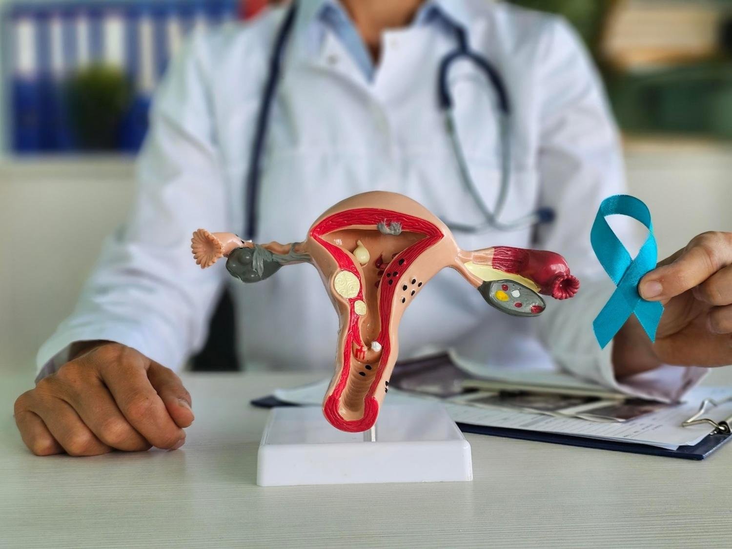
Gestational Trophoblastic Disease
GTD — a group of rare diseases in which abnormal trophoblast cells grow inside the uterus after conception
Gestational Trophoblastic Disease
GTD — a group of rare diseases in which abnormal trophoblast cells grow inside the uterus after conception
Fast Facts
- Gestational trophoblastic disease (GTD) is a group of rare diseases in which abnormal trophoblast cells grow inside the uterus after conception.
- GTD can be treated and in the majority of cases the treatment results in a cure.
- Most women who have had a single incidence of GTD can go on to have normal pregnancies.
- Hydatidiform mole (HM) is the most common type of GTD.
- Signs of GTD include abnormal vaginal bleeding and a uterus that is larger than normal.
About GTD
Gestational trophoblastic disease (GTD) is a general term for rare tumors that form from the tissues surrounding a fertilized egg. Instead of a healthy fetus developing, a tumor forms. The tissue is made of trophoblast cells and normally surrounds the fertilized egg in the uterus. Trophoblast cells help connect the fertilized egg to the wall of the uterus and form part of the placenta (the organ that passes nutrients from the mother to the fetus). Until there are signs or symptoms of the tumor, the pregnancy will seem like a normal pregnancy.
GTD is often found early and usually cured. More than 80 percent of GTD cases are non-cancerous. Hydatidiform mole (HM) is the most common type of GTD. Most women who have had a single incidence of GTD can go on to have normal pregnancies.
Most GTD is benign (not cancer) and does not spread, but some types become malignant (cancer) and spread to nearby tissues or distant parts of the body.
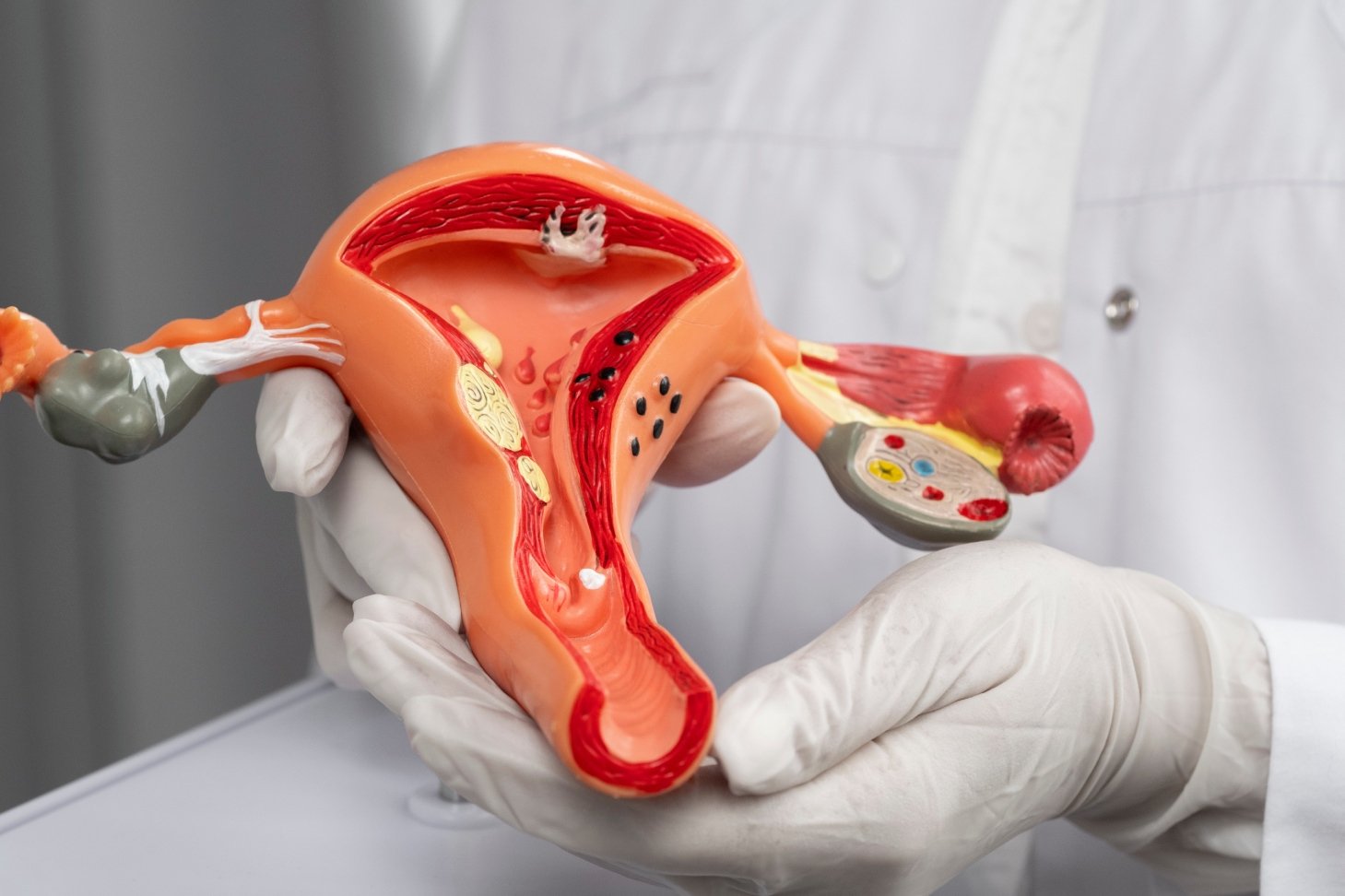
Hydatidiform mole
- A hydatidiform mole is typically a benign, treatable condition. This rare, slow-growing tumor arises after the sperm and egg join and do not develop into a fetus; instead, tissue that resembles grape-like cysts forms. Hydatidiform mole is the most common type of gestational trophoblastic tumor. It is also called molar pregnancy. It is usually benign (not cancer) but it may spread to nearby tissues (invasive mole).
- This disease generally becomes evident six to ten weeks after conception—usually when a woman, believing that she is pregnant, starts having vaginal bleeding. Around six to ten weeks of pregnancy is also when a woman typically begins her prenatal care with an obstetrician and has her first ultrasound and blood work; this is the point at which most women are diagnosed with a hydatiform mole.
- A woman with a hydatiform mole will register a positive home urine pregnancy test because these kits detect the presence of a hormone produced during pregnancy called beta-human beta-chorionic gonadotropin (beta-hCG, or HCG). However, the level of HCG, measured with a blood test at a doctor’s office, can be higher in women with a hydatiform mole than in women experiencing a normal pregnancy.
Choriocarcinoma
A choriocarcinoma is even rarer than a hydatidiform mole. This type of GTD may have begun as a hydatidiform mole or may arise from tissue that remains in the uterus following a miscarriage or full-term delivery of a baby.
Unlike a hydatidiform mole, a choriocarcinoma is a malignant and more aggressive form of GTD that spreads into the muscle wall of the uterus. A choriocarcinoma can also spread more widely to other parts of the body such as the lungs, liver, and/or brain.
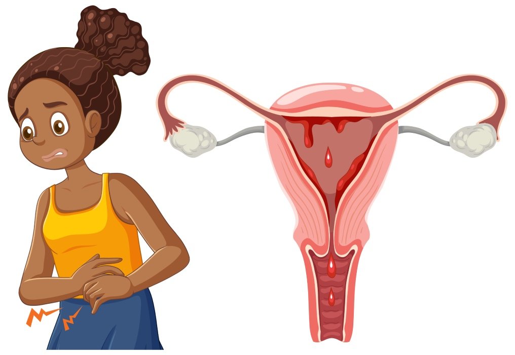
Signs and Symptoms
A woman with an hydatidiform mole (partial or complete) or choriocarcinoma may experience one or more of these symptoms:
- irregular, non-menstrual vaginal bleeding, possibly with blood clots or a watery brown discharge
- pelvic pain or discomfort
- nausea and vomiting that are more frequent and severe than what a woman typically experiences during a normal pregnancy
- fatigue and shortness of breath due to anemia resulting from blood loss through vaginal bleeding
- faster growth than expected for weeks of pregnancy, due to extension of the uterus
- rapid heartbeat, warm skin, and mild tremor or shaking; caused by an overactive thyroid gland, this complication may occur rarely in women with high HCG levels
- preeclampsia (also known as toxemia)—a pregnancy-related condition which can cause a sharp rise in blood pressure
Risk Factors
Although some studies have linked GTD (molar pregnancy) with dietary or genetic factors, the real cause is still unknown.
GTD appears to be more common at the beginning and end of the reproductive age group. Thus, women becoming pregnant younger than 20 or older than 35 years of age are at higher risk.
Those who have a personal history of hydatidiform mole are also at higher risk of having a second molar pregnancy.
Testing and Detection
Pelvic exam: By examination the doctor may find that the uterus is larger than normal for the duration (age) of the pregnancy.
Ultrasound examination of the pelvis: An ultrasound examination will make the diagnosis and will help distinguish mole from other early pregnancy abnormalities.
Blood tests: For GTD, the blood is checked for the level of beta human chorionic gonadotropin (beta-hCG), a hormone that is made by the body during pregnancy. Beta-hCG in the blood of a woman who is not pregnant may be a sign of GTD.
Urinalysis: A test to check the color of urine and its contents, such as sugar, protein, blood, bacteria, and the level of beta-hCG.
Treatment
The doctor may remove the cancer using one of the following operations:
Dilatation and curettage (D&C) with suction evacuation: A surgical procedure to remove abnormal tissue and parts of the inner lining of the uterus. The cervix is dilated and the material inside the uterus is removed with a small vacuum-like device. The walls of the uterus are then gently scraped with a curette (spoon-shaped instrument) to remove any material that may remain in the uterus.
Hysterectomy: Surgery to remove the uterus, and sometimes the cervix.
After the doctor removes all the cancer that can be seen at the time of the surgery, some patients may be given chemotherapy after surgery to kill any cancer cells that are left.
Chemotherapy
Chemotherapy is a cancer treatment that uses drugs to stop the growth of cancer cells, either by killing the cells or by stopping them from dividing. When chemotherapy is taken by mouth or injected into a vein or muscle, the drugs enter the bloodstream and can reach cancer cells throughout the body (systemic chemotherapy). When chemotherapy is placed directly into the cerebrospinal fluid, an organ, or a body cavity such as the abdomen, the drugs mainly affect cancer cells in those areas (regional chemotherapy). The way the chemotherapy is given depends on the type and stage of the cancer being treated, or whether the tumor is low-risk or high-risk.
Combination chemotherapy is treatment using more than one anticancer drug.
Radiation therapy
Radiation therapy is a cancer treatment that uses high-energy x-rays or other types of radiation to kill cancer cells or keep them from growing. There are two types of radiation therapy:
External radiation therapy uses a machine outside the body to send radiation toward the cancer.
Internal radiation therapy uses a radioactive substance sealed in needles, seeds, wires, or catheters that are placed directly into or near the cancer.
The way the radiation therapy is given depends on the type of gestational trophoblastic disease being treated. External radiation therapy is used to treat gestational trophoblastic disease.